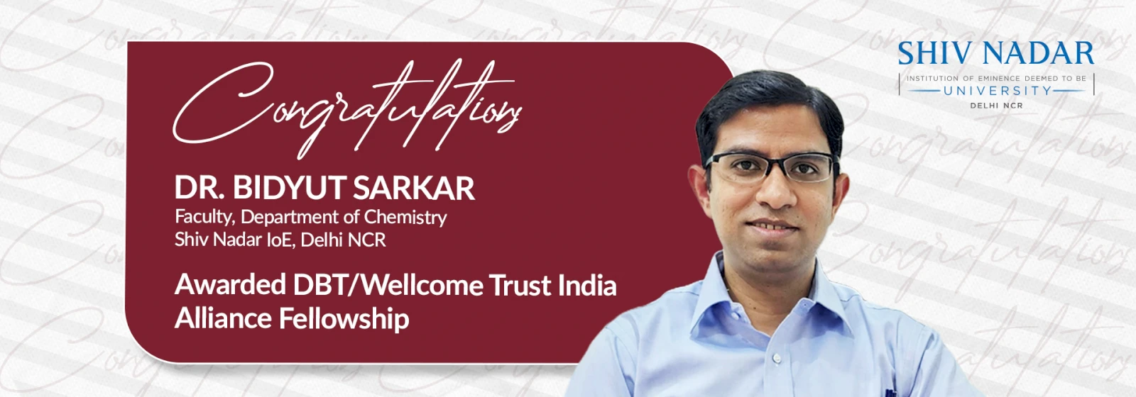
Dr. Bidyut Sarkar from the Department of Chemistry has been awarded the DBT/Wellcome Trust India Alliance Fellowship.
From a scientific perspective, understanding life entails understanding its underlying biochemical processes, and the biological machinery that makes everything we know possible. Over the past few decades, our knowledge of cell biology has skyrocketed thanks to the development of novel microscopy and analytical techniques, with no signs of slowing down.
The Department of Chemistry at the School of Natural Sciences, Shiv Nadar University (SNU), Delhi-NCR is making substantial contributions to research in this field through the work of Dr. Bidyut Sarkar—an emerging leader in the fields of fluorescence spectroscopy and microscopy applied to biochemistry and biophysics research. In July 2023, Dr. Sarkar was awarded the DBT/Wellcome Trust India Alliance Intermediate Fellowship to conduct studies at SNU in an emerging and exciting area: how non-coding RNAs regulate genetic expression at the single-molecule level.
Interestingly, Dr. Sarkar’s academic journey was not initially close to this topic. While pursuing his master’s and Ph.D. degrees in organic and biophysical chemistry between 2006 and 2014, Dr. Sarkar was captivated by the use of cutting-edge fluorescence and optical spectroscopy techniques in biophysical research. Driven by a desire to invest his efforts in answering medically relevant questions, he dove into studying the biophysics of amyloid-beta protein aggregation, which occurs in diseases like Alzheimer’s. He also focused on developing a novel label-free imaging technique to visualise neurotransmitters at the single-cell level.
His immersion in these topics sparked an interest in the precise details of how biomacromolecules work, including the dynamic relationship that exists between a molecule’s structure and its biological functions. He spent nine years studying abroad at RIKEN, Japan, advancing and utilising a new technique called 2D Fluorescence Lifetime Correlation Spectroscopy (2D FLCS), which was originally developed in his postdoctoral laboratory in RIKEN. “With 2D FLCS, one can look into structural changes of single biomacromolecules at time resolutions in the order of microseconds or less than a microsecond, which is approximately the speed limit at which proteins can fold,” explains Dr. Sarkar.
During his time in Japan, he used this state-of-the-art method to shed light on how non-coding RNA in bacteria affect gene expression. However, noticing that very few countries were conducting studies using this technology, Dr. Sarkar decided to return to India to expand his home country’s horizons into this mostly uncharted scientific territory. Seeking funding, he applied for the DBT/Wellcome Trust India Alliance Intermediate Fellowship.
His efforts paid off, and Dr. Sarkar is currently undertaking two major research projects at SNU under this Fellowship. The first focuses on riboswitches, a type of non-coding RNA that bind cellular metabolites to regulate subsequent RNA transcription and protein translation. Since riboswitches are present in bacteria but not in human cells, they constitute potential antibiotic targets with minimal side effects. Theoretically, one could design alternative ligands that bind to riboswitches, altering or interrupting their function and ultimately killing bacteria. In practice, however, this approach has not been very successful thus far, despite efforts during the past few years. “I hypothesize that the ligand must make changes to the riboswitch at a sufficiently fast speed because the transcriptional regulation through riboswitches happen right as the downstream nascent RNA chain is being produced,” posits Dr. Sarkar. “This is where the dynamics aspect of the ligand binding comes in. Thus, I want to look into the structural changes of these non-coding RNA, as well as their folding/unfolding kinetics upon interaction with the ligand. With this molecular-level understanding, it might be possible to not only propose new drugs that work better, but also understand why they work.”
His second project explores the interference mechanisms of microRNAs, which can cause gene silencing. Unlike small interfering RNAs, that are highly specific and silence only a single target mRNA, microRNAs are more versatile and can have hundreds of potential targets. Thus, their silencing mechanisms are much less known. Focusing on the regulatory effects of microRNA obtained from influenza virus, Dr. Sarkar plans to design and build a one-of-a-kind modular experimental setup combining single-molecule spectroscopic techniques (such as 2D FLCS) and fluorescence-based transcriptomics and proteomics. This will enable him to observe the structural dynamics of microRNAs at the single-molecule level, covering timescale from a microsecond to about one minute, and study their regulatory effects on cellular transcriptome and proteome.
Notably, these cutting-edge techniques are relatively low-cost compared to other analytical tools and capture more information. “Unlike RNA sequencing, there is no need to amplify the signal; moreover, the spatial information is not lost, since we are not dealing with a cellular extract,” remarks Dr. Sarkar. “One of my dreams would be to use the results we get from looking at the molecular picture in collaboration with pharmaceutical chemists to develop novel or more effective drugs.”
We sincerely wish Dr. Sarkar and colleagues the best of luck in current and future endeavours. May their research efforts lead to breakthroughs in biology and medicine!
