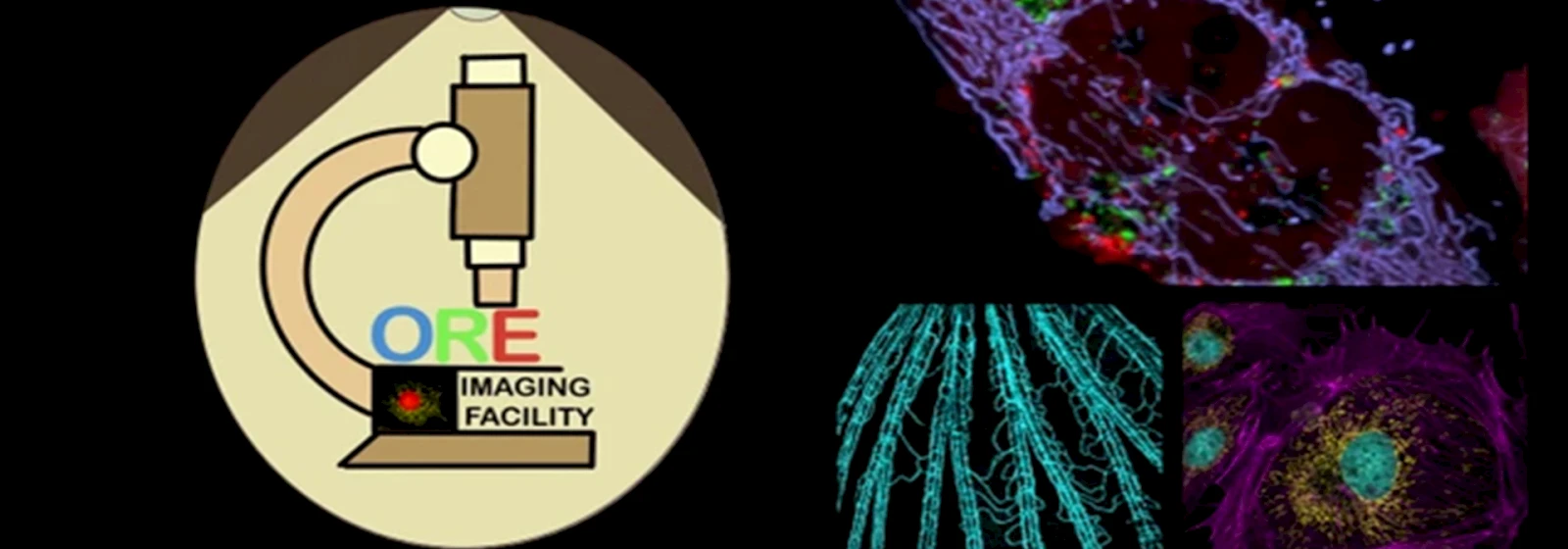
Core Imaging Facility (CIF)
Overview
The Core Imaging Facility (CIF) at the Department of Life Sciences, School of Natural Sciences, Shiv Nadar IoE University, Delhi-NCR, is dedicated to providing cutting-edge imaging solutions for advanced research in life sciences. Equipped with state-of-the-art microscopy systems, including confocal and fluorescence imaging, the facility serves as a hub for interdisciplinary collaboration, innovation, and technical excellence. Our mission is to support and enhance the research capabilities of scientists, fostering groundbreaking discoveries across a wide spectrum of biological sciences.
Vision
Our vision is to establish the Core Imaging Facility as a globally recognized center for imaging excellence. We aim to lead in the integration of advanced imaging technologies with life science research, empowering scientists to explore and understand complex biological systems with unprecedented precision. Through innovation, collaboration, and continuous development, we strive to push the boundaries of what is possible in biological imaging, contributing to impactful scientific discoveries that benefit society at large.
Mission
- To provide world-class imaging support to researchers, enabling high-quality data acquisition and analysis through state-of-the-art instrumentation.
- To foster a collaborative environment where interdisciplinary research thrives, promoting knowledge-sharing and technological advancements in imaging techniques.
- To continuously upgrade and expand our imaging capabilities, ensuring that researchers have access to the latest innovations in microscopy and imaging analysis.
- To train the next generation of scientists in advanced imaging techniques, empowering them with the skills needed for cutting-edge research.
- To serve as a regional and national resource, partnering with institutions and industry to address critical challenges in life sciences through advanced imaging solutions.
Available Instruments
- Laser Scanning Confocal Microscope: A1R MP ready with Ti2E Microscope, Nikon (DST-FIST Funded Instrument)
The coherent light (excitation laser) passes through a pinhole aperture that is situated in a conjugate plane (confocal) with a scanning point on the specimen and a second pinhole aperture positioned in front of the detector (a photomultiplier tube). As the laser is reflected by a dichromatic mirror and scanned across the specimen in a defined focal plane, the emission from points on the specimen (in the same focal plane) passes back through the dichromatic mirror and is focused as a confocal point at the detector pinhole aperture. Due to this aperture, the detector does not detect the extraneous light (out of focus), resulting in sharp images.
About Microscope: The Microscope consists of an inverted fluorescence research microscope (Ti2E) with the following objectives:
- 2X Plan Apochromat (NA 0.10)
- 10X Plan Apochromat (NA 0.45)
- 20X Plan Apochromat (NA 0.75)
- 40X Plan-Apochromat DIC (NA 0.95)
- 60X Plan-Apochromat Oil immersion DIC (NA 1.40)
- 100X Plan-Apochromat Oil immersion DIC (NA 1.40)
Observation and imaging units: A Mercury Lamp (Intensilight C-HGFIE)-) illuminator for sample observation and four lasers for confocal imaging.
Technical Information about the system:
- Lasers: We have solid-state lasers, and lines are 405/488/561/640nm
- Minimum Pixel Set: 32X32
- Maximum Pixel Set: 4096X4096
- Maximum Frame Rate: 420 Frames per Second (512X32 pixels) Galvano scanner and Resonant scanner.
- As regards PFS it is 30fps @ 512x 512 in Resonant Scanner and 420fps@512x 32
- We have Tokai Hit live-cell imaging setup with a perfect focus system (PFS).
Detection unit:
- Transmitted light detector T-PMT
- LSM A1R MP, Nikon confocal with Galvano, and resonant scanner head with 2MA-PMT and 2 GaAsP (for spectral detection)
Applications: Fluorescence imaging of tissues and cells, live imaging, spectral imaging, and dynamics experiments such as FRAP and FRET.
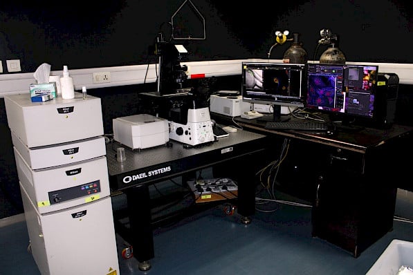
Nikon, A1R MP Ti2E Confocal Microscope (DST-FIST Funded )
- Laser Scanning Confocal Microscope: AXR NSPARC with Ti2E Microscope, Nikon
The coherent light (excitation laser) passes through a pinhole aperture that is situated in a conjugate plane (confocal) with a scanning point on the specimen and a second pinhole aperture positioned in front of the detector (a photomultiplier tube). As the laser is reflected by a dichromatic mirror and scanned across the specimen in a defined focal plane, the emission from points on the specimen (in the same focal plane) passes back through the dichromatic mirror and is focused as a confocal point at the detector pinhole aperture. Due to this aperture, the detector does not detect the extraneous light (out of focus), resulting in sharp images.
About Microscope: The Microscope consists of an inverted fluorescence research microscope (Ti2E) with the following objectives:
- 4X Plan Apochromat (NA 0.21)
- 10X Plan Apochromat (NA 0.45)
- 20X Plan Apochromat (NA 0.80)
- 40X Plan-Apochromat DIC (NA 0.95)
- 60X Plan-Apochromat Oil immersion DIC (NA 1.42)
- 100X Plan-Apochromat Oil immersion DIC (NA 1.45)
Observation and imaging units: A Nikon D-LEDI illuminator for sample observation and four lasers for confocal imaging.
Technical Information about the system:
- Lasers: We have solid-state lasers, and lines are 405/488/561/640nm
- Scanner Type : Hybrid ( Galvano and Resonant )
- FOV: 25mm for Both Galvano and Resonant scanner.
- Minimum Pixel Set: 2k x 16 ( Resonant scanner)
- Maximum Pixel Set: 8192 x 8192 ( Galvano Scanner) & 2k x 2k ( Resonant Scanner)
- Frame Rate: a) Galvano Scanner : 1-2 fps @ 512 x 512 and 10fps @ 512x 512 with 8x zoom and maximum 240fps @ 512x 16
- Frame Rate : Resonant Scanner : 30fps @ 2k x 512 and maximum 720fps @ 512x 16
- Live cell Imaging : Available from Tokai Hit live-cell imaging setup with a perfect focus system (PFS).
Detection unit:
Transmitted light detector T-PMT
4 GaAsP detector for standard confocal imaging for 4 color simultaneous imaging and Spectral resolution is 5nm with 1nm Tunability .
Additional Super Resolution Detector NSPARC, which is capable to achieve Lateral resolution appr. 100nm and Axial resolution of 300nm , its is compatible with both Galvano and Resonant Scanner .
Applications: Fluorescence imaging of tissues, model Animals and cells, live imaging, spectral imaging, and dynamics experiments such as FRAP and FRET.
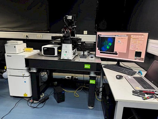
Nikon, AXR NSPARC Ti2E Confocal Microscope
- Fluorescence Microscope: Ti E Microscope (Nikon, Microscope)
About Microscope: The Microscope consists of an inverted fluorescence research microscope (Ti E) that can be exploited for both fixed cell imaging in the fluorescence mode as well as fixed stage-position live cell imaging. Illumination is through a halogen lamp and detection through a CCD camera, controlled by the NIS Elements software suite with the following objectives:
- 10X Plan Apochromat (NA 0.45)
- 20X Plan Apochromat (NA 0.75)
- 40X Plan-Apochromat Oil immersion DIC (NA 0.95)
- 60X Plan-Apochromat Oil immersion DIC (NA 1.40)
- 100X Plan-Apochromat Oil immersion DIC (NA 1.40)
Applications: Fluorescence imaging of tissues, model Animals and cells, live imaging.
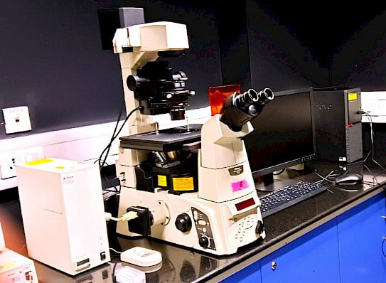
Nikon, TiE Fluorescence Microscope
Capabilities of the microscope:
Most of the systems are in inverted microscopes, having the full range of lasers with the following capabilities.
- Fluorescence recovery after photobleaching (FRAP)
- Fluorescence resonance energy transfer (FRET)
- 3-D Imaging & reconstruction
- Live-cell Imaging
- Morphological studies of cells & tissue
- Photo-bleaching investigations
- DNA Hybridization
- Membrance dynamic & ion probes
- Drug discovery
- Lambda scan
Services:
- Optical Microscope
- Image Analysis
- Microscope Training
Outreach:
- Microscope Training
- Course workshops
- Knowledge exchange
- Access to cutting-edge technologies
Publications
- Kalarikkal C, Anjali, Bhattacharjee S, Mapa K, P CAS. Lipid droplet specific BODIPY based rotors with viscosity sensitivity to distinguish normal and cancer cells: impact of molecular conformation. J Mater Chem B. 2025 Jan 22;13(4):1474-1486. doi: 10.1039/d4tb02405b. PMID: 39698835.
- Mondal A, Mukherjee S, Upadhyay P, Saxena I, Pati S, Singh S. Enhancing NADPH to restore redox homeostasis and lysosomal function in G6PD-deficient microglia. Heliyon. 2025 Feb 15;11(4):e42735. doi: 10.1016/j.heliyon.2025.e42735. PMID: 40084013; PMCID: PMC11903804.
- Mondal A, Mukherjee S, Upadhyay P, Saxena I, Pati S, Singh S. Enhancing NADPH to restore redox homeostasis and lysosomal function in G6PD-deficient microglia. Heliyon. 2025 Feb 15;11(4):e42735. doi: 10.1016/j.heliyon.2025.e42735. PMID: 40084013; PMCID: PMC11903804.
- Modi M, Chauhan D, Gilmore MC, Cava F, Priyadarshini R. Deficiency in peptidoglycan recycling promotes β-lactam sensitivity in Caulobacter crescentus. mBio. 2025 Apr 9;16(4):e0297524. doi: 10.1128/mbio.02975-24. Epub 2025 Mar 11. PMID: 40066998; PMCID: PMC11980594.
- Kumar V, Chauhan L, Singh D, Kumar A, Kulandaisamy R, Kushwaha T, Baswal K, Singh R, Kumar S, Gholap SL, Hariprasad P, Dadinaboyina SB, Thota JR, Sehgal D, Appaiahgari MB, Inampudi KK. Ethyl acetate extract of Ruta graveolens: a specific and potent inhibitor against the drug-resistant EGFR_T790M mutant in NSCLC. Front Pharmacol. 2025 Apr 29;16:1570108. doi: 10.3389/fphar.2025.1570108. PMID: 40365309; PMCID: PMC12069052.
- Modi M, Chauhan D, Gilmore MC, Cava F, Priyadarshini R. Deficiency in peptidoglycan recycling promotes β-lactam sensitivity in Caulobacter crescentus. mBio. 2025 Apr 9;16(4):e0297524. doi: 10.1128/mbio.02975-24. Epub 2025 Mar 11. PMID: 40066998; PMCID: PMC11980594.
- Gupta H, Singh A, Gupta A. Cancer-associated mutation at glycine 400 in TIP60 disrupt its phase separation property and catalytic activity resulting in compromised DNA damage repair function of the cell. Biochem Biophys Res Commun. 2025 Mar 19;753:151457. doi: 10.1016/j.bbrc.2025.151457. Epub 2025 Feb 6. PMID: 39965267.
- Lawrence M, Khurana J, Gupta A. Identification, characterization, and CADD analysis of Plasmodium DMAP1 reveals it as a potential molecular target for new anti-malarial discovery. J Biomol Struct Dyn. 2025 May;43(8):4258-4273. doi: 10.1080/07391102.2024.2302923. Epub 2024 Jan 12. PMID: 38217317.
- Saroj S, Saha S, Ali A, Gupta SK, Bharadwaj A, Agrawal T, Pal S, Rakshit T. Plant Extracellular Nanovesicle-Loaded Hydrogel for Topical Antibacterial Wound Healing In Vivo. ACS Appl Bio Mater. 2025 Jan 20;8(1):1-11. doi: 10.1021/acsabm.4c00992. Epub 2024 Oct 8. PMID: 39377525.
- Pareek N, Kalita N, Pandey R, Samanta A. Methionine-Derived Fluorescent Probes Targeting Mitochondria: A Tool for Real-Time Oxidative Stress Monitoring in Live Cells. Chembiochem. 2025 Feb 3;26(5):e202400893. doi: 10.1002/cbic.202400893. Epub 2025 Jan 20. PMID: 39797544.
- Pareek N, Yadav R, Munan S, Baruah M, Samanta A. A Fluorescent Probe Differentiates Apoptosis from Cysteine-Deprivation Ferroptosis through Imaging of Viscosity and Lipid Droplets. Chemistry. 2025 Mar 3;31(13):e202404523. doi: 10.1002/chem.202404523. Epub 2025 Jan 13. PMID: 39751778.
- Tiwari A, Lee SJ, Thokchom AK. Surfactant-based interface capture towards the development of 2D-printed photonic structures. Mater Horiz. 2025 Apr 14;12(8):2689-2700. doi: 10.1039/d4mh01560f. PMID: 39831825.
- Pareek N, Kalita N, Pandey R, Samanta A. Methionine-Derived Fluorescent Probes Targeting Mitochondria: A Tool for Real-Time Oxidative Stress Monitoring in Live Cells. Chembiochem. 2025 Feb 3;26(5):e202400893. doi: 10.1002/cbic.202400893. Epub 2025 Jan 20. PMID: 39797544.
- Pareek N, Yadav R, Munan S, Baruah M, Samanta A. A Fluorescent Probe Differentiates Apoptosis from Cysteine-Deprivation Ferroptosis through Imaging of Viscosity and Lipid Droplets. Chemistry. 2025 Mar 3;31(13):e202404523. doi: 10.1002/chem.202404523. Epub 2025 Jan 13. PMID: 39751778.
- Biligiri KK, Sharma NR, Mohanty A, Sarkar DP, Vemula PK, Rampalli S. A cytoplasmic form of EHMT1N methylates viral proteins to enable inclusion body maturation and efficient viral replication. PLoS Biol. 2024 Nov 7;22(11):e3002871. doi: 10.1371/journal.pbio.3002871. PMID: 39509467; PMCID: PMC11575796.
- Saroj S, Saha S, Ali A, Gupta SK, Bharadwaj A, Agrawal T, Pal S, Rakshit T. Plant Extracellular Nanovesicle-Loaded Hydrogel for Topical Antibacterial Wound Healing In Vivo. ACS Appl Bio Mater. 2025 Jan 20;8(1):1-11. doi: 10.1021/acsabm.4c00992. Epub 2024 Oct 8. PMID: 39377525.
- Nagar, T. Agrawal, T. Rakshit, B., Dhar Catalyst Free Synthesis of 4-Aryl/Alkylamino-1,2-Naphthoquinones: Applications in Fluorescence Microscopy * Chemistry Select. 2024. DOI: 10.1002/slct.202402531.
- Munan S, Kottarathil S, Joseph MM, Jana A, Ali M, Mapa K, Maiti KK, Samanta A. IndiFluors: A New Full-Visible Color-Tunable Donor-Acceptor-Donor (D1-A-D2) Fluorophore Family for Ratiometric pH Imaging during Mitophagy. ACS Sens. 2024 Jul 26;9(7):3502-3510. doi: 10.1021/acssensors.1c02381. Epub 2022 Feb 3. PMID: 35113517.
- Jha MP, Kumar V, Ghosh A, Mapa K. Sse1, Hsp110 chaperone of yeast, controls the cellular fate during endoplasmic reticulum stress. G3 (Bethesda). 2024 Jun 5;14(6):jkae075. doi: 10.1093/g3journal/jkae075. PMID: 38577891; PMCID: PMC11152076.
- Haider S, Chakraborty S, Chowdhury G, Chakrabarty A. Opposing Interplay between Nuclear Factor Erythroid 2-Related Factor 2 and Forkhead BoxO 1/3 is Responsible for Sepantronium Bromide's Poor Efficacy and Resistance in Cancer cells: Opportunity for Combination Therapy in Triple Negative Breast Cancer. ACS Pharmacol Transl Sci. 2024 Apr 29;7(5):1237-1251. doi: 10.1021/acsptsci.3c00279. PMID: 38751638; PMCID: PMC11091984.
- Shakeel I, Haider S, Khan S, Ahmed S, Hussain A, Alajmi MF, Chakrabarty A, Afzal M, Imtaiyaz Hassan M. Thymoquinone, artemisinin, and thymol attenuate proliferation of lung cancer cells as Sphingosine kinase 1 inhibitors. Biomed Pharmacother. 2024 Aug;177:117123. doi: 10.1016/j.biopha.2024.117123. Epub 2024 Jul 14. PMID: 39004062.
- Chakraborty S, Haider S, Mukherjee G, Chakrabarty A, Chowdhury G. O6-Alkylguanine-DNA Alkyltransferase Maintains Genome Integrity by Forming DNA-Protein Cross-Links during Inflammation-Associated Peroxynitrite-Mediated DNA Damage. Chem Res Toxicol. 2024 Dec 16;37(12):1952-1964. doi: 10.1021/acs.chemrestox.4c00296. Epub 2024 Oct 21. PMID: 39431584.
- Mondal A, Munan S, Saxena I, Mukherjee S, Upadhyay P, Gupta N, Dar W, Samanta A, Singh S, Pati S. G6PD deficiency mediated impairment of iNOS and lysosomal acidification affecting phagocytotic clearance in microglia in response to SARS-CoV-2. Biochim Biophys Acta Mol Basis Dis. 2024 Oct;1870(7):167444. doi: 10.1016/j.bbadis.2024.167444. Epub 2024 Jul 27. PMID: 39074627.
- Munan S, Mondal A, Shailja S, Pati S, Samanta A. Unique Synthetic Strategy for Probing in Situ Lysosomal NO for Screening Neuroinflammatory Phenotypes against SARS-CoV-2 RNA in Phagocytotic Microglia. Anal Chem. 2024 May 14;96(19):7479-7486. doi: 10.1021/acs.analchem.3c05981. Epub 2024 May 1. PMID: 38689560.
- Prajapat SK, Mishra L, Khera S, Owusu SD, Ahuja K, Sharma P, Choudhary E, Chhabra S, Kumar N, Singh R, Kaushal PS, Mahajan D, Banerjee A, Motiani RK, Vrati S, Kalia M. Methotrimeprazine is a neuroprotective antiviral in JEV infection via adaptive ER stress and autophagy. EMBO Mol Med. 2024 Jan;16(1):185-217. doi: 10.1038/s44321-023-00014-w. Epub 2024 Jan 2. PMID: 38177535; PMCID: PMC10897192.
- Yadav R, Munan S, Ali M, Mapa K, Samanta A. A Systematic Design and Synthesis of PET-based Fluorescent Probes for Monitoring pH During Mitophagy. Chem Asian J. 2023 Jun 15;18(12):e202300308. doi: 10.1002/asia.202300308. Epub 2023 May 11. PMID: 37126645.
- Niranjan R, Zafar S, Lochab B, Priyadarshini R. Synthesis and Characterization of Sulfur and Sulfur-Selenium Nanoparticles Loaded on Reduced Graphene Oxide and Their Antibacterial Activity against Gram-Positive Pathogens. Nanomaterials (Basel). 2022 Jan 7;12(2):191. doi: 10.3390/nano12020191. PMID: 35055210; PMCID: PMC8782023.
- Kumar A, Hooda P, Puri A, Khatter R, S Al-Dosari M, Sinha N, Parvez MK, Sehgal D. Methotrexate, an anti-inflammatory drug, inhibits Hepatitis E viral replication. J Enzyme Inhib Med Chem. 2023 Dec;38(1):2280500. doi: 10.1080/14756366.2023.2280500. Epub 2023 Nov 17. PMID: 37975328; PMCID: PMC11003484.
- Hooda P, Al-Dosari M, Sinha N, Parvez MK, Sehgal D. Inhibition of HEV Replication by FDA-Approved RdRp Inhibitors. ACS Omega. 2023 Oct 27;8(44):41570-41578. doi: 10.1021/acsomega.3c05637. PMID: 37969986; PMCID: PMC10633873.
- Ali A, Saroj S, Saha S, Gupta SK, Rakshit T, Pal S. Glucose-Responsive Chitosan Nanoparticle/Poly(vinyl alcohol) Hydrogels for Sustained Insulin Release In Vivo. ACS Appl Mater Interfaces. 2023 Jul 12;15(27):32240-32250. doi: 10.1021/acsami.3c05031. Epub 2023 Jun 27. PMID: 37368956.
- Biswas S, Baruah M, Shil A, Sarkar S, Ali M, Samanta A, Bhuniya S. Polarity-Driven Two-Photon Fluorescent Probe for Monitoring the Perturbation in Lipid Droplet Levels during Mitochondrial Dysfunction and Acute Pancreatitis. ACS Sens. 2023 Oct 27;8(10):3793-3803. doi: 10.1021/acssensors.3c01245. Epub 2023 Oct 10. PMID: 37815484.
- Baruah M, Jana A, Pareek N, Singh S, Samanta A. A Ratiometric Fluorescent Probe for Hypochlorite and Lipid Droplets to Monitor Oxidative Stress. Biosensors (Basel). 2023 Jun 17;13(6):662. doi: 10.3390/bios13060662. PMID: 37367027; PMCID: PMC10295842.
- Maiti D, Munan S, Singh S, Das R, Samanta A, Sen S. Light induced diversity-oriented synthesis (DOS) library of annulated indolizine fluorophores for imaging non-lysosomal lipid droplets (LDs). J Mater Chem B. 2023 Mar 8;11(10):2191-2199. doi: 10.1039/d2tb02656b. PMID: 36779938.
- Sarkar R, Rao KBN, Jha MP, Mapa K. Endoplasmic reticulum-unfolded protein response pathway modulates the cellular response to mitochondrial proteotoxic stress. Cell Stress Chaperones. 2022 May;27(3):241-256. doi: 10.1007/s12192-022-01264-2. Epub 2022 Mar 16. PMID: 35294718; PMCID: PMC9106787.
- Chakraborty S, Mallick D, Goswami M, Guengerich FP, Chakrabarty A, Chowdhury G. The Natural Products Withaferin A and Withanone from the Medicinal Herb Withania somniferaAre Covalent Inhibitors of the SARS-CoV-2 Main Protease. J Nat Prod. 2022 Oct 28;85(10):2340-2350. doi: 10.1021/acs.jnatprod.2c00521. Epub 2022 Sep 13. PMID: 36098617; PMCID: PMC9491402.
- Chakrabarti A, Kaushik M, Khan J, Soota D, Ponnusamy K, Saini S, Manvati S, Singhal J, Ranganathan A, Pati S, Dhar PK, Singh S. tREPs-A New Class of Functional tRNA-Encoded Peptides. ACS Omega. 2022 May 25;7(22):18361-18373. doi: 10.1021/acsomega.2c00661. PMID: 35694484; PMCID: PMC9178612.
- Anam Z, Kumari G, Mukherjee S, Rex DAB, Biswas S, Maurya P, Ravikumar S, Gupta N, Kushawaha AK, Sah RK, Chaurasiya A, Singhal J, Singh N, Kaushik S, Prasad TSK, Pati S, Ranganathan A, Singh S. Complementary crosstalk between palmitoylation and phosphorylation events in MTIP regulates its role during Plasmodium falciparum Front Cell Infect Microbiol. 2022 Sep 29;12:924424. doi: 10.3389/fcimb.2022.924424. PMID: 36250062; PMCID: PMC9556994.
- Chakrabarti A, Narayana C, Joshi N, Garg S, Garg LC, Ranganathan A, Sagar R, Pati S, Singh S. Metalloprotease Gp63-Targeting Novel Glycoside Exhibits Potential Antileishmanial Activity. Front Cell Infect Microbiol. 2022 May 4;12:803048. doi: 10.3389/fcimb.2022.803048. PMID: 35601095; PMCID: PMC9115111.
- Soni S, Singh D, Aggarwal R, Veerapu NS. Enhanced fitness of hepatitis C virus increases resistance to direct-acting antivirals. J Gen Virol. 2022 Feb;103(2). doi: 10.1099/jgv.0.001699. PMID: 35133954.
- Hooda P, Chaudhary M, Parvez MK, Sinha N, Sehgal D. Inhibition of Hepatitis E Virus Replication by Novel Inhibitor Targeting Methyltransferase. Viruses. 2022 Aug 15;14(8):1778. doi: 10.3390/v14081778. PMID: 36016400; PMCID: PMC9415367.
- Mishra VS, Patil S, Reddy PC, Lochab B. Combinatorial delivery of CPI444 and vatalanib loaded on PEGylated graphene oxide as an effective nanoformulation to target glioblastoma multiforme: In vitro Front Oncol. 2022 Aug 16;12:953098. doi: 10.3389/fonc.2022.953098. PMID: 36052261; PMCID: PMC9426685.
- Patil S, Mishra VS, Yadav N, Reddy PC, Lochab B. Dendrimer-Functionalized Nanodiamonds as Safe and Efficient Drug Carriers for Cancer Therapy: Nucleus Penetrating Nanoparticles. ACS Appl Bio Mater. 2022 Jul 18;5(7):3438-3451. doi: 10.1021/acsabm.2c00373. Epub 2022 Jun 27. PMID: 35754387.
- Dubey S, Jaiswal B, Gupta A. TIP60 acts as a regulator of genes involved in filopodia formation and cell migration during wound healing. J Biol Chem. 2022 Jul;298(7):102015. doi: 10.1016/j.jbc.2022.102015. Epub 2022 May 5. PMID: 35525269; PMCID: PMC9249863.
- Jaiswal B, Agarwal A, Gupta A. Lysine Acetyltransferases and Their Role in AR Signaling and Prostate Cancer. Front Endocrinol (Lausanne). 2022 Aug 17;13:886594. doi: 10.3389/fendo.2022.886594. PMID: 36060957; PMCID: PMC9428678.
- Saxena H, Gupta A. Plasmodium falciparum PfRUVBL proteins bind at the TARE region and var gene promoter located in the subtelomeric region. Pathog Dis. 2022 Jul 21;80(1):ftac018. doi: 10.1093/femspd/ftac018. PMID: 35640888.
- Paul D, Paul A, Mukherjee D, Saroj S, Ghosal M, Pal S, Senapati D, Chakrabarti J, Pal SK, Rakshit T. A Mechanoelastic Glimpse on Hyaluronan-Coated Extracellular Vesicles. J Phys Chem Lett. 2022 Sep 15;13(36):8564-8572. doi: 10.1021/acs.jpclett.2c01629. Epub 2022 Sep 7. PMID: 36069730.
- Shivappagowdar A, Garg S, Srivastava A, Hada RS, Kalia I, Singh AP, Garg LC, Pati S, Singh S. Pathogenic Pore Forming Proteins of PlasmodiumTriggers the Necrosis of Endothelial Cells Attributed to Malaria Severity. Toxins (Basel). 2021 Jan 15;13(1):62. doi: 10.3390/toxins13010062. PMID: 33467515; PMCID: PMC7839052.
- Pal P, Modi M, Ravichandran S, Yennamalli RM, Priyadarshini R. DNA-Binding Properties of YbaB, a Putative Nucleoid-Associated Protein From Caulobacter crescentus. Front Microbiol. 2021 Oct 28;12:733344. doi: 10.3389/fmicb.2021.733344. PMID: 34777284; PMCID: PMC8581549.
- Yadav N, Monisha M, Niranjan R, Dubey A, Patil S, Priyadarshini R, Lochab B. Antibacterial performance of fully biobased chitosan-grafted-polybenzoxazine films: Elaboration and properties of released material. Carbohydr Polym. 2021 Feb 15;254:117296. doi: 10.1016/j.carbpol.2020.117296. Epub 2020 Oct 24. PMID: 33357864.
Patents Details
- Title: A Lysosome-Specific Nitric Oxide Probe for Neuroinflammation and Process of Synthesis Thereof
- Inventors: Samanta, A., Munan, S., Pati, S., Mondal, A., Singh, S.
- Patent Filed: Application No. 202311040064, dated June 12, 2023
- Title: A Lysosome-Specific Nitric Oxide Probe for Neuroinflammation and Process of Synthesis Thereof
- Inventors: Abir Mondal, Subrata Munan, Shailja Singh*, Soumya Pati*, Animesh Samanta* (*Main Inventor)
- Patent Published: Application No. 202311040064 A, published on July 21, 2023
- Title: Glycoside-Based Anti-Toxin for Epsilon Intoxication and Preparation Method Thereof
- Inventors: Chintam Narayana, Abhishek Shivappagowdar, Ram Sagar, Soumya Pati*, Shailja Singh* (*Main Inventor)
- Patent Published: Application No. 201811034958 A, published in The Patent Office Journal No. 12/2020, dated March 20, 2020.
External Users: We have more than 30 institutions with external users, some of them listed below.
- AIIMS Delhi
- JNU Delhi
- IGIB New Delhi
- RCB Faridabad
- THSTI Faridabad
- IISER Pune
- Jamia Milia Islamia Delhi
- NIT (Calicut)
- InStem Bangalore
Recognition of the Facility
The facility is a Member of the Indian BioImaging (IBI) Committee and the Global BioImaging (GBI)
Contact:
Core Imaging Facility (CIF)- [email protected]
R Block's second floor (Room no R250 & R-248) and B Block's first floor (Imaging Facility Room at CoEE), Department of Life Sciences, SNS. Shiv Nadar IoE University, Noida, Delhi-NCR
Mr. Rajan Singh (Facility-In-Charge) – [email protected]
Oversees the Core Imaging Facility, Committee
Dr. Rudra Nayan Das (CIF, Faculty-In-Charge) – [email protected]
Dr. Koyeli Mapa (CIF, Committee Member)- [email protected]
Dr. Richa Priyadarshini (CIF, Committee Member)- [email protected]
Statement of Acknowledgement (for DST-FIST Confocal Microscope)
“This research work was carried out in part at the DST-FIST Confocal Microscope Facility (R-250), a part of the Core Imaging Facility (CIF) of the Department of Life Sciences, SNS, which is managed by the Shiv Nadar IoE DTU Delhi-NCR and is funded by the Department of Science and Technology (Grant No. SR/FST/LS-1/2017/59(c)).”
Name of the instrument- Nikon Confocal laser scanning microscope
[Model: Nikon Ti2E, A1R MP (multiphoton ready) with motorized inverted microscope]
Statement of Acknowledgement (for Nikon AXR- NSPARC Confocal Microscope)
“This research work was carried out at the Core Imaging Facility (CIF) of the Department of Life Sciences, SNS, managed by the Shiv Nadar IoE DTU Delhi-NCR, and is funded by the Core SN IoE University Delhi-NCR funding.
Name of the instrument- Nikon Confocal laser scanning microscope
[Model: Nikon Ti2E, AXR NSPARC (with a motorized inverted microscope)]
microscope]